Wednesday 20 June 2012

Human anatomy of head and neck

The skull is made up of 19 bones, 12 of which are pairs. You
can see most of these bones in the illustration. Where the
bones join is called a suture. Little fingers of bone interdigitate
with adjoining little fingers to make the joining solid.
The bones shown are: the frontal bone, the nasal bones, the
lacrimal bones, the ethmoid bones, the sphenoid bones, the
zygomatic bones, the maxilla, the mandible, the parietal
bones, the temporal bones, the occipital bone, the palatine
bones (not seen, since they are inside the orbit of the eye),
and the vomer (not seen, since it is inside the nasal cavity).

There are two bones that you cannot see in the illustration,
the palatine and the vomer. The palatine bones are paired
and are buried deep in the skull behind the nose. They make
up the rear part of the palate, part of the base of the nasal
cavity, and a small part of the floor or the orbit. The vomer is
a thin bone which forms part of the nasal septum separating
the two sides of the nasal cavity.

Lacrimal, meaning tear producing, is from the Latin lachrymal,
meaning a small vase, of the kind found in ancient
Roman sepulchers that was used for collecting tears shed in

mourning.(The lacrimal bone forms half of the receptacle,
which holds the lacrimal sac, a structure that receives the
tears and directs them into the nasal cavity. That explains
why we blow our noses in cold weather, or when we cry, we
are blowing out the tears that have drained into the cavity.
The other half of the receptacle for the lacrimal sac is made
from the frontal process of the maxilla.) Ethmoid, so-named
because it is full of holes, is from the Greek ethmo and oiedes,
meaning “formed like a strainer.” Sphenoid is from the Greek
spheno and eidos together meaning wedge-shaped. Zygomatic
or zygoma comes from the Greek zygon, which means yoke,
the kind used to harness oxen. Maxilla is from the Latin mala
meaning jaw, particularly the upper jaw. Mandible derives
from the Latin mandibula, which stems from mandare meaning
to chew and pertains particularly to the lower jaw, which
has most of the chewing motion. Parietal is from the Latin
paries, parietes, meaning “walls of a hollow cavity.” Temporal
indicates the temple, from the Latin tempora, meaning “temple,
the right place, the fatal spot..”(As well as indicating a
place on the skull where death can easily be afflicted, this
word coveys a sense of reverence for life.) Occipital is from
the Latin occiput, meaning “the back of the head.” Palatine is
from the Latin palatum, meaning the hard palate and is the
base for the words palatable and palliative. Vomer is the Latin
word for plowshare.

Search term
Human anatomy of head and neck


Tuesday 19 June 2012

what is myology? The defination of myology

The study of muscles is called myology after the Greek word mys, which means “mouse.”
Muscles perform a number of functions vital to maintaining life, including

Movement: Skeletal muscles (those attached to bones) convert chemical energy into
mechanical work, producing movement ranging from finger tapping to a swift kick of a
ball by contracting, or shortening. Reflex muscle reactions protect your fingers when
you put them too close to a fire and startle you into watchfulness when an unexpected
noise sounds. Many purposeful movements require several sets, or groups, of muscles
to work in unison.

Vital functions: Without muscle activity, you die. Muscles are doing their job when
your heart beats, when your blood vessels constrict, and when your intestines squeeze
food along your digestive tract in peristalsis.

Antigravity: Perhaps that’s overstating it, but muscles do make it possible for you to
stand and move about in spite of gravity’s ceaseless pull. Did your mother tell you to
improve your posture? Just think how bad it would be without any muscles!

Heat generation: You shiver when you’re cold and stamp your feet and jog in place
when you need to warm up. That’s because chemical reactions in muscles result in
heat, helping to maintain the body’s temperature.

Keep the body together: Muscles are the warp and woof of your body’s structure,
binding one part to another.

As you may remember from studying tissues, muscle cells — called fibers — are some
of the longest in the body. Fibers are held together by connective tissue and enclosed
in a fibrous sheath called fascia. Some muscle fibers contract rapidly, whereas others
move at a leisurely pace. Generally speaking, however, the smaller the structure to be
moved, the faster the muscle action. Exercise can increase the thickness of muscle
fibers, but it doesn’t make new fibers. Skeletal muscles have a rich vascular supply
that dilates during exercise to give the working muscle the extra oxygen it needs to
keep going.

Two processes are central to muscle development in the developing embryo: myogenesis,
during which muscle tissue is formed; and morphogenesis, when the muscles form
into internal organs. By the eighth week of gestation, a fetus is capable of coordinated
movement.

Search terms;
what is myology
what the myology
mylogy
defination of myology

Arthrology, The Defination of Arthrology

Arthrology, which stems from the ancient Greek word arthros (meaning “jointed”), is
the study of those structures that hold bones together, allowing them to move to varying
degrees — or fixing them in place — depending on the design and function of the
joint. The term articulation, or joint, applies to any union of bones, whether it moves
freely or not at all.

Inside some joints, such as knees and elbows, are fluid-filled sacs called bursae that
help reduce friction between tendons and bones; inflammation in these sacs is called
bursitis. Some joints are stabilized by connective tissue called ligaments that range
from bundles of collagenous fibers that restrict movement and hold a joint in place to
elastic fibers that can repeatedly stretch and return to their original shapes.

The three types of joints are as follows:


Fibrous: Fibrous tissue rigidly joins the bones in a form of articulation called
synarthrosis, which is characterized by no movement at all. The sutures of the
skull are fibrous joints.

Cartilaginous: This type of joint is found in two forms:

• Synchondrosis articulation involves rigid cartilage that allows no movement,
such as the joint between the ribs, costal cartilage, and sternum.
• Symphysis joints occur where cartilage fuses bones in such a way that
pressure can cause slight movement, called amphiarthrosis. Examples
include the intervertebral discs and the symphysis pubis.

Synovial: Also known as diarthrosis, or freely moving, joints, this type of articulation
involves a synovial cavity, which contains articular fluid secreted from the
synovial membrane to lubricate the opposing surfaces of bone. The synovial
membrane is covered by a fibrous joint capsule layer that’s continuous with the
periosteum of the bone. Ligaments surrounding the joint strengthen the capsule
and hold the bones in place, preventing dislocation. In some synovial joints,
such as the knee, fibrous connective tissue called meniscus develops in the
cavity, dividing it into two parts. In the knee, this meniscus stabilizes the joint
and acts as a shock absorber.

What is muscle tissue made up of

The question in your minds about what is muscle tissue made up of.in histology you should know that muscle tissue is made up of fibers known as myocytes. The cytoplasm within the fibers
is called sarcoplasm, and within that sarcoplasm are minute myofibrils that contain the
protein filaments actin and myosin. These filaments slide past each other during a
muscle contraction, shortening the fiber.

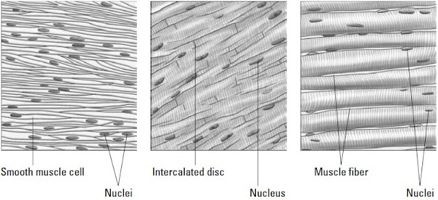
Following are the three types of muscle tissue

Smooth muscle tissue: This type of tissue contracts without conscious control.
Made up of spindle-shaped fibers with large, centrally located nuclei, it’s found in
the walls of internal organs, or viscera. Smooth muscle gets its name from the
fact that, unlike other muscle tissue types, it is not striated.

Cardiac muscle tissue: Also known as myocardium, cardiac muscle tissue is
made of branching fibers, each with a central nucleus and alternating light and
dark striations. Between the fibers are dark structures called intercalated discs.
As with smooth muscle, cardiac muscle tissue contractions occur through the
autonomic nervous system.

Skeletal, or striated, muscle tissue: Biceps, triceps, pecs — these are the muscles
that bodybuilders focus on. As the name implies, skeletal muscles attach to
the skeleton and are used throughout the central nervous system for movement.
Muscle fibers are cylindrical with several nuclei in each cell (which makes them
multinucleated) and cross-striations throughout.


What is muscle tissue made up of


“ Take A Journey Inside The Mysterious
      World Of The Human Body ... ”

This heavily illustrated self-teaching course
gives you everything you need to -
CLICK HERE


Monday 18 June 2012

Get under your skin definition

Perhaps because of its unique job of both protecting the outer body and lining internal
organs, epithelial tissue comes in more varieties than any other tissue.

Epithelial tissues, which generally are arranged in sheets or tubes of tightly-packed cells,
always have a free, or apical, surface that can be exposed to the air or to fluid. That free surface
also can be covered by additional layers of epithelial tissue. But whether it’s layered or
not, each epithelial cell has polarity, and all but one side of the cell is
tucked snugly against neighboring cells. The apical side sometimes has cytoplasmic projections
such as cilia, hair-like growths that can move material over the cell’s surface, or
microvilli, finger-like projections that increase the cell’s surface area for absorption. Opposite
the apical side is the basal side (think basement), which typically attaches to some kind of
connective tissue.

Epithelial tissue serves several key functions, including the following:

Protection: Skin protects vulnerable structures or tissues deeper in the body.

Barrier: Epithelial tissues prevent foreign materials from getting inside the body.

Sensation: Sensory nerve endings embedded in epithelial tissue connect the
body with outside stimuli.

Secretion: Epithelial tissue in glands can be specialized to secrete enzymes, hormones,
and fluids.

Single-layer epithelial tissue is classified as simple. Tissue with more than one layer is
called stratified. Epithelial tissues also can be classified according to shape: Squamous
is a thin, flat cell; cuboidal is, as the name implies, equal in height and width and
shaped like a cube; and columnar cells are taller than they are wide.