Tuesday 24 July 2012

what is cardiovascular system function

Today i am talk about "what is cardiovascular system function"
Moving to the Beat of a Pump Also called the cardiovascular system, 
the circulatory system includes the heart, all blood
vessels, and the blood that moves endlessly through it all . It’s what’s
referred to as a closed double system; the term “closed” is used for three reasons: because
the blood is contained in the heart and its vessels; because the vessels specifically target the
blood to the tissues; and because the heart critically regulates blood flow to the tissues.
The system is called “double” because there are two distinct circuits and cavities within the
heart separated by a wall of muscle called the septum. (Each cavity in turn has two chambers
called atria on top and ventricles below). The double circuits are the following:

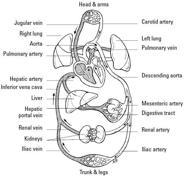
1. The pulmonary circuit carries blood to and from the lungs for gaseous exchange.
Centered in the right side of the heart, this circuit receives blood saturated with
carbon dioxide from the veins and pumps it through the pulmonary artery (or trunk)
to capillaries in the lungs, where the carbon dioxide departs the system. That same
blood, freshly loaded with oxygen, then returns to the left side of the heart through
the pulmonary veins where it enters the second circuit.

2. The systemic circuit uses the oxygen-rich blood to maintain a constant internal environment
around the body’s tissues. From the left side of the heart, the blood moves
through the aorta to a variety of systemic arteries for distribution throughout the body.

After oxygen is exchanged for carbon dioxide, the blood returns to the pulmonary
circuit on the right side of the heart via the superior and inferior venae
cavae (the singular is vena cava).

Although cutely depicted in popular culture as uniformly curvaceous, the heart actually
looks more like a blunt, muscular cone (roughly the size of a fist) resting on the
diaphragm. A fluid-filled, fibrous sac called the pericardium (or heart sac) wraps
loosely around the package; it’s attached to the large blood vessels emerging from the
heart but not to the heart itself. The sternum (breastbone) and third to sixth costal
cartilages of the ribs provide protection in front of (ventrally to) the heart. Behind it lie
the fifth to eighth thoracic vertebrae. Two-thirds of the heart lies to the left of the
body’s center, with its apex (cone) pointed down and to the left. At less than 5 inches
long and a bit more than 3 inches wide, an adult human heart weighs around 10
ounces — a couple ounces shy of a can of soda.

Three layers make up the wall of the heart.

1. On the outside lies the epicardium (or visceral pericardium), which is composed
of fibroelastic connective tissue dappled with adipose tissue (fat) that fills external
grooves called sulci (the singular is sulcus). The larger coronary vessels and
nerves are found in the adipose tissue that fills the sulci.

2. Beneath the epicardium lies the myocardium, which is composed of layers and
bundles of cardiac muscle tissue.

3. The endocardium, the heart’s interior lining, is composed of simple squamous
endothelial cells.

Too much to remember? To keep the layers straight, turn to the Greek roots. Epi– is
the Greek term for “upon” or “on” whereas endo– comes from the Greek endon meaning
“within.” The medical definition of myo– is “muscle.” And peri– comes from the
Greek term for “around” or “surround.” Hence the epicardium is on the heart, the
endocardium is inside the heart, the myocardium is the muscle between the two,
and the pericardium surrounds the whole package. By the way, the root cardi– comes
from the Greek word for heart, kardia.

The pericardium is made up of two parts — a tough inelastic sac called the fibrous pericardium
on the outside and a serous (or lubricated) membrane nearer the heart called
the parietal pericardium. Between the serous layers of the epicardium and the parietal
pericardium is the small pericardial space and its tiny amount of lubricating pericardial
fluid. This watery substance prevents irritation during systole (contraction of the
heart) and diastole (relaxation of the heart).


you may also search for:
function of cardiovascular system
cardiovascular system function
human cardiovascular system
cardiovascular system heart
anatomy physiology cardiovascular system heart functions

Monday 23 July 2012

What is inside the lungs

what is inside the lungs,After the pharynx and larynx comes the 
trachea, more popularly known as the windpipe.
Roughly 6 inches long in adults, it’s a tube connected to the larynx in front of the
esophagus that’s made up of C-shaped rings of hyaline cartilage and fibrous connective
tissue that strengthen it and keep it open. Like the larynx, the trachea’s lined with
mucous membrane covered in cilia. Just above the heart, the trachea splits into two
bronchi divided by a sharp ridge called the carina, with each leading to a lung. But
they’re not identical: The right primary bronchus is shorter and wider than the left primary
bronchus. Each primary bronchus divides into secondary bronchi with a branch
going to each lobe of the lung; the right side gets three secondary bronchi while the
left gets only two. Once inside a designated lobe, the bronchus divides again into tertiary
bronchi. The right lung has ten such branches: three in the superior (or upper)
lobe, two in the middle lobe, and five in the inferior (or lower) lobe. The left lung has
only four tertiary bronchi: two in the upper lobe and two in the lower lobe.

Each tertiary bronchi subdivides one more time into smaller tubes called bronchioles
which lack the supporting cartilage of the larger structures. Each
bronchiole ends in an elongated sac called the atrium (also known as an alveolar duct
or alveolar sac). Alveoli (or air cells) surround the atria, as do small capillaries that
pick up oxygen for delivery elsewhere in the body and dump off carbon dioxide
fetched from elsewhere. Overall, there are 23 branches in the respiratory system, with
a combined surface area (counting the alveoli) the size of a tennis court!

Knowing that the bronchi aren’t evenly distributed, you may have guessed that the
lungs aren’t identical either. You’re right. They’re both spongy and porous because of
the air in the sacs, but the right lung is larger, wider, and shorter than the left lung and
has three lobes. The left lung divides into only two lobes and is both narrower and
longer to make room for the heart because two-thirds of that organ lies to the left of
the body’s midline. Each lobe is made up of many lobules, each with a bronchiole
ending in an atrium inside.

Covering each lung is a thin serous membrane called the visceral pleura that folds back
on itself to form a second outer layer, the parietal pleura, with a pleural cavity between
the two layers. These two layers secrete a watery fluid into the cavity to lubricate the
surfaces that rub against each other as you breathe. When the pleural membrane
becomes inflamed in a condition called pleurisy, a sticky discharge roughens the
pleura, causing painful irritation. An accompanying bacterial infection means that pus
accumulates in the pleural cavity in a condition known as empyema.


Blood comes to the lungs through two sources: the pulmonary arteries and the
bronchial arteries. The pulmonary trunk comes from the right ventricle of the heart
and then branches into the two pulmonary arteries carrying venous blood (the only
arteries that contain blood loaded with carbon dioxide from various parts of the body)
to the lungs. That blood goes through capillaries in the lungs where the carbon dioxide
leaves the blood and enters the alveoli to be expelled during exhalation; oxygen leaves
the alveoli through the capillaries to enter the bloodstream. After that, oxygenated
arterial blood returns to the left atrium through the pulmonary veins (the only veins
that contain oxygenated blood), completing the cycle. Bronchial arteries branch off
the thoracic aorta of the heart, supplying the lung tissue with nutrients and oxygen.

"What is inside the lungs"

Sweat glands in human body


Humans perspire over nearly every inch of skin, but anyone with sweaty palms or
smelly feet can attest to the fact that sweat glands are most numerous in the palms
and soles, with the forehead running a close third. There are two types of sweat, or
sudoriferous, glands: eccrine and apocrine. Both are coiled tubules embedded in the
dermis or subcutaneous layer composed of simple columnar cells.

Eccrine glands are distributed widely over the body — an average adult has roughly
3 million of them — and produce the watery, salty secretion you know as sweat. Each
gland’s duct passes through the epidermis to the skin’s surface, where it opens as a
sweat pore. The sympathetic division of the autonomic nervous system controls when

and how much perspiration is secreted depending on how hot the body becomes.
Sweat helps cool the skin’s surface by evaporating as fast as it forms. About 99 percent
of eccrine-type sweat is water, but the remaining 1 percent is a mixture of sodium chloride
and other salts, uric acid, urea, amino acids, ammonia, sugar, lactic acid, and
ascorbic acid.

Apocrine sweat glands are located primarily in armpits (known as the axillary region)
and the groin area. Usually associated with hair follicles, they produce a white, cloudy
secretion that contains organic matter. Although apocrine-type sweat contains the
same basic components as eccrine sweat and also is odorless when first secreted, bacteria
quickly begin to break down its additional fatty acids and proteins — explaining
the post-exercise underarm stench. In addition to exercise, sexual and other emotional
stimuli can cause contraction of cells around these glands, releasing sweat.

Thursday 19 July 2012

Structure of human brain and function


The human brain is a soft, gelatinous collection of gray and white matter
encased in the cranium and weighing about 1,400 grams (roughly three pounds)
in the adult. Estimates vary, but there may be 100 billion or more neurons in the
brain, and at least ten times this number of glial cells.As an indicator of the 
astonishing  degree of connectivity between cerebral neurons, each one 
makes contact with as  many as 10,000 others Interneurons, situated 
between afferent and efferent neurons, constitute by far the largest class 
of brain neurons, so that the great majority of the brain’s neuronal activity
is concerned with the processing and transfer of information that occur 
between sensory input and motor output In other words, a large quantity 
of nervous tissue lies interposed between the sensory and motor systems
 to elaborate the phenomena of behavior.

The brain is made up of the cerebrum, the brainstem, and the cerebellum
Most important for the higher functions is the cerebrum, which comprises
 the paired cerebral hemispheres and the diencephalon, the main 
components of which are the thalamus and hypothalamus. Why the 
hemispheres are paired, and why they have distinct functional 
affiliations in contrast to other paired organs in the body such as the 
lungs and kidneys, are not understood, but the distinct operations of 
the two cerebral hemispheres will be frequently emphasized in this book. 
The hemispheres are folded into ridges called gyri, and the grooves 
between these are known as sulci or fissures. These gross neuroanatomical 
features form the basis for the division of the hemispheres into four lobes: frontal,
temporal, parietal, and occipital.

The parcellation of the hemispheres into four lobes is somewhat arbitrary
but serves to produce convenient neuroanatomical landmarks that have important
functional affiliations. The image above gives a brief outline of some prominent
brain-behavior relationships, which will be developed in greater detail throughout
this book. The frontal lobes, largest and most anterior, provide the origin
of the motor system via the corticospinal tracts, mediate the production of language
and prosody, and organize the integrative capacities of motivation, comportment,
and executive function. The temporal lobes receive primary auditory
input, mediate comprehension of language and prosody, and, in concert with
the closely connected limbic system, subserve important aspects of memory
and emotion. The parietal lobes receive tactile input, mediate visuospatial competence,
and subserve reading and calculation skills. The occipital lobes, smallest
and most posterior, receive primary visual input and mediate perception of
visual material before further processing occurs in more anterior regions.

"Structure of human brain and function"
                                 

Tuesday 17 July 2012

Epidermis Layers


Epidermis, which contains no blood vessels, is made up of layers of closely packed
epithelial cells. From the outside in, these layers are the following:

Stratum corneum epidermis layers (literally the “horny layer”) is about 20 layers of flat, scaly,dead cells containing a type of water-repellent protein called keratin. These cells, which represent about three-quarters of the thickness of the epidermis, are said to be cornified, which means that they’re tough and horny like the cells that form hair or fingernails. Humans shed this layer of tough, durable skin at a prodigious rate; in fact, much of household dust consists of these flaked-off cells. Where the skin is rubbed or pressed more often, cell division increases, resulting in calluses and corns.

Stratum lucidum epidermis layers (from the Latin word for “clear”) is found only
in the thick skin on the palms of the hands and the soles of the feet. This translucent layer of
dead cells contains eleidin, a protein that becomes keratin as the cells migrate
into the stratum corneum, and it consists of cells that have lost their nuclei and
cytoplasm.

Stratum granulosum epidermis layers is three to five layers of flattened cells containing keratohyalin,a substance that marks the beginning of keratin formation. No nourishment
from blood vessels reaches this far into the epidermis, so cells are either
dead or dying by the time they reach the stratum granulosum. The nuclei of cells
found in this layer are degenerating; when the nuclei break down entirely, the
cell can’t metabolize nutrients and dies.

Stratum spinosum epidermis layers(also sometimes called the spinous layer) has ten layers containing prickle cells, named for the spine-like projections that connect them with
other cells in the layer. Langerhans cells, believed to be involved in the body’s
immune response, are prevalent in the upper portions of this layer and sometimes
the lower part of the stratum granulosum; they migrate from the skin to
the lymph nodes in response to infection. Some mitosis (cell division) takes
place in the stratum spinosum, but the cells lose the ability to divide as they
mature.

Stratum basale epidermis layers(or stratum germinativum) is also referred to as the germinal layer
because this single layer of mostly columnar stem cells generates all the cells
found in the other epidermal layers. It rests on the papillary (rough or bumpy)
surface of the dermis, close to the blood supply needed for nourishment and
oxygen. The mitosis that constantly occurs here replenishes the skin; it takes
about two weeks for the cells that originate here to migrate up to the stratum
corneum, and it’s another two weeks before they’re shed. About a quarter of this
layer’s cells are melanocytes, cells that synthesize a pale yellow to black pigment
called melanin that contributes to skin color and provides protection against
ultraviolet radiation (the kind of radiation found in sunlight). The remaining
cells in this layer become keratinocytes, the primary epithelial cell of the skin.
Melanocytes secrete melanin directly into the keratinocytes in a process called
cytocrine secretion. Merkel’s cells, a large oval cell believed to be involved in the
sense of touch, occasionally appear amid the keratinocytes.

In addition to melanin, the epidermis contains a yellowish pigment called carotene (the
same one found in carrots and sweet potatoes). Found in the stratum corneum and the
fatty layers beneath the skin, it produces the yellowish hue associated with Asian
ancestry or increased carrot consumption. The pink to red color of Caucasian skin is
caused by hemoglobin, the red pigment of the blood cells. Because Caucasian skin contains
relatively less melanin, hemoglobin can be seen more easily through the epidermis.
Sometimes the limited melanin in Caucasian skin pools in small patches. Can you
guess the name of those patches of color? Yep, they’re freckles. Albinos, on the other
hand, have no melanin in their skin at all, making them particularly sensitive to ultraviolet
radiation.


Ridges and grooves form on the outer surface of the epidermis to increase the friction
needed to grasp objects or move across slick surfaces. On hands and feet, these ridges
form patterns of loops and whorls — fingerprints, palm prints, and footprints — that
are unique to each person. You leave these imprints on smooth surfaces because of
the oily secretions of the sweat glands on the skin’s surface. In addition to these finer
patterns, the areas around joints develop patterns called flexion lines. Deeper and
more permanent lines are called flexion creases.

About tissues in the human body

Today we talk about tissues in the human body. Muscle tissue is classified in three ways based on the tissue’s function, shape, and structure:

Smooth muscle tissue: So-called because it doesn’t have the cross-striations typical
of other kinds of muscle, the spindle-shaped fibers of smooth muscle tissue
do have faint longitudinal striping. This muscle tissue forms into sheets and
makes up the walls of hollow organs such as the stomach, intestines, and bladder.
The tissue’s involuntary movements are relatively slow, so contractions last
longer than those of other muscle tissue, and fatigue is rare. Each fiber is about
6 microns in diameter and can vary from 15 microns to 500 microns long. If
arranged in a circle inside an organ, contraction constricts the cavity inside the
organ. If arranged lengthwise, contraction of smooth muscle tissue shortens
the organ.

Cardiac muscle tissue: Found only in the heart, cardiac muscle fibers are
branched, cross-striated, feature one central nucleus, and move through involuntary
control. An electron microscope view of the tissue shows separate fibers
tightly pressed against each other, forming cellular junctions called intercalated
discs that look like tiny, dark-colored plates. Some experts believe intercalated
discs are not cellular junctions but rather special structures that help move an
electrical impulse throughout the heart.

Skeletal muscle tissue:
This is the tissue that most people think of as muscle.
It’s the only muscle subject to voluntary control through the central nervous
system. Its long, striated cylindrical fibers contract quickly but tire just as fast.
Skeletal muscle, which is also what’s considered meat in animals, is 20 percent
protein, 75 percent water, and 5 percent organic and inorganic materials. Each
multinucleated fiber is encased in a thin, transparent membrane called a sarcolemma
that receives and conducts stimuli. The fibers, which vary from 10
microns to 100 microns in diameter and up to 4 centimeters in length, are subdivided
lengthwise into tiny myofibrils roughly 1 micron in diameter that are suspended
in the cell’s sarcoplasm.

Now you know about tissues in the human body


tissues of the human body
types of tissues in human body
tissue in human body
how many tissues are in the human body

Monday 9 July 2012

Human being digestive system


Before jumping into a discussion on the alimentary tract, we need to review some basic
terms.


Ingestion: Taking in food

Digestion: Changing the composition of food — splitting large molecules into smaller
ones — to make it usable by the cells

Deglutition: Swallowing, or moving food from the mouth to the stomach

Absorption: Occurs when digested food moves through the intestinal wall and into
the blood

Egestion: Eliminating waste materials or undigested foods at the lower end of the
digestive tract; also known as defecation


The alimentary tract develops early on in a growing embryo. The primitive gut, or archenteron,
develops from the endoderm (inner germinal layer) during the third week after conception,
a stage during which the embryo is known as a gastrula. At the anterior end (head end),
the oral cavity, nasal passages, and salivary glands develop from a small depression called
a stomodaeum in the ectoderm (outer germinal layer). The anal and urogenital structures
develop at the opposite, or posterior, end from a depression in the ectoderm called the
proctodaeum. In other words, the digestive tract develops from an endodermal tube with
ectoderm at each end.

Whereas the respiratory tract is a two-way street — oxygen flows in and carbon dioxide flows
out — the digestive tract is designed to have a one-way flow (although when you’re sick or
your body detects something bad in the food you’ve eaten, what goes down sometimes comes
back up).


Mouth Pharynx Esophagus Stomach Small intestine Large intestine

When you swallow food, it’s mixed with digestive enzymes in both saliva and stomach
acids. Circular muscles on the inside of the tract and long muscles along the outside of
the tract keep the material moving right through defecation at the end of the line.


Search terms:

process digestion human beings
digestive system of human body
digestive system of man