Tuesday, 24 July 2012

what is cardiovascular system function

Today i am talk about "what is cardiovascular system function"
Moving to the Beat of a Pump Also called the cardiovascular system, 
the circulatory system includes the heart, all blood
vessels, and the blood that moves endlessly through it all . It’s what’s
referred to as a closed double system; the term “closed” is used for three reasons: because
the blood is contained in the heart and its vessels; because the vessels specifically target the
blood to the tissues; and because the heart critically regulates blood flow to the tissues.
The system is called “double” because there are two distinct circuits and cavities within the
heart separated by a wall of muscle called the septum. (Each cavity in turn has two chambers
called atria on top and ventricles below). The double circuits are the following:

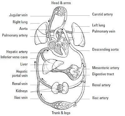
1. The pulmonary circuit carries blood to and from the lungs for gaseous exchange.
Centered in the right side of the heart, this circuit receives blood saturated with
carbon dioxide from the veins and pumps it through the pulmonary artery (or trunk)
to capillaries in the lungs, where the carbon dioxide departs the system. That same
blood, freshly loaded with oxygen, then returns to the left side of the heart through
the pulmonary veins where it enters the second circuit.

2. The systemic circuit uses the oxygen-rich blood to maintain a constant internal environment
around the body’s tissues. From the left side of the heart, the blood moves
through the aorta to a variety of systemic arteries for distribution throughout the body.

After oxygen is exchanged for carbon dioxide, the blood returns to the pulmonary
circuit on the right side of the heart via the superior and inferior venae
cavae (the singular is vena cava).

Although cutely depicted in popular culture as uniformly curvaceous, the heart actually
looks more like a blunt, muscular cone (roughly the size of a fist) resting on the
diaphragm. A fluid-filled, fibrous sac called the pericardium (or heart sac) wraps
loosely around the package; it’s attached to the large blood vessels emerging from the
heart but not to the heart itself. The sternum (breastbone) and third to sixth costal
cartilages of the ribs provide protection in front of (ventrally to) the heart. Behind it lie
the fifth to eighth thoracic vertebrae. Two-thirds of the heart lies to the left of the
body’s center, with its apex (cone) pointed down and to the left. At less than 5 inches
long and a bit more than 3 inches wide, an adult human heart weighs around 10
ounces — a couple ounces shy of a can of soda.

Three layers make up the wall of the heart.

1. On the outside lies the epicardium (or visceral pericardium), which is composed
of fibroelastic connective tissue dappled with adipose tissue (fat) that fills external
grooves called sulci (the singular is sulcus). The larger coronary vessels and
nerves are found in the adipose tissue that fills the sulci.

2. Beneath the epicardium lies the myocardium, which is composed of layers and
bundles of cardiac muscle tissue.

3. The endocardium, the heart’s interior lining, is composed of simple squamous
endothelial cells.

Too much to remember? To keep the layers straight, turn to the Greek roots. Epi– is
the Greek term for “upon” or “on” whereas endo– comes from the Greek endon meaning
“within.” The medical definition of myo– is “muscle.” And peri– comes from the
Greek term for “around” or “surround.” Hence the epicardium is on the heart, the
endocardium is inside the heart, the myocardium is the muscle between the two,
and the pericardium surrounds the whole package. By the way, the root cardi– comes
from the Greek word for heart, kardia.

The pericardium is made up of two parts — a tough inelastic sac called the fibrous pericardium
on the outside and a serous (or lubricated) membrane nearer the heart called
the parietal pericardium. Between the serous layers of the epicardium and the parietal
pericardium is the small pericardial space and its tiny amount of lubricating pericardial
fluid. This watery substance prevents irritation during systole (contraction of the
heart) and diastole (relaxation of the heart).


you may also search for:
function of cardiovascular system
cardiovascular system function
human cardiovascular system
cardiovascular system heart
anatomy physiology cardiovascular system heart functions

Monday, 23 July 2012

What is inside the lungs

what is inside the lungs,After the pharynx and larynx comes the 
trachea, more popularly known as the windpipe.
Roughly 6 inches long in adults, it’s a tube connected to the larynx in front of the
esophagus that’s made up of C-shaped rings of hyaline cartilage and fibrous connective
tissue that strengthen it and keep it open. Like the larynx, the trachea’s lined with
mucous membrane covered in cilia. Just above the heart, the trachea splits into two
bronchi divided by a sharp ridge called the carina, with each leading to a lung. But
they’re not identical: The right primary bronchus is shorter and wider than the left primary
bronchus. Each primary bronchus divides into secondary bronchi with a branch
going to each lobe of the lung; the right side gets three secondary bronchi while the
left gets only two. Once inside a designated lobe, the bronchus divides again into tertiary
bronchi. The right lung has ten such branches: three in the superior (or upper)
lobe, two in the middle lobe, and five in the inferior (or lower) lobe. The left lung has
only four tertiary bronchi: two in the upper lobe and two in the lower lobe.

Each tertiary bronchi subdivides one more time into smaller tubes called bronchioles
which lack the supporting cartilage of the larger structures. Each
bronchiole ends in an elongated sac called the atrium (also known as an alveolar duct
or alveolar sac). Alveoli (or air cells) surround the atria, as do small capillaries that
pick up oxygen for delivery elsewhere in the body and dump off carbon dioxide
fetched from elsewhere. Overall, there are 23 branches in the respiratory system, with
a combined surface area (counting the alveoli) the size of a tennis court!

Knowing that the bronchi aren’t evenly distributed, you may have guessed that the
lungs aren’t identical either. You’re right. They’re both spongy and porous because of
the air in the sacs, but the right lung is larger, wider, and shorter than the left lung and
has three lobes. The left lung divides into only two lobes and is both narrower and
longer to make room for the heart because two-thirds of that organ lies to the left of
the body’s midline. Each lobe is made up of many lobules, each with a bronchiole
ending in an atrium inside.

Covering each lung is a thin serous membrane called the visceral pleura that folds back
on itself to form a second outer layer, the parietal pleura, with a pleural cavity between
the two layers. These two layers secrete a watery fluid into the cavity to lubricate the
surfaces that rub against each other as you breathe. When the pleural membrane
becomes inflamed in a condition called pleurisy, a sticky discharge roughens the
pleura, causing painful irritation. An accompanying bacterial infection means that pus
accumulates in the pleural cavity in a condition known as empyema.


Blood comes to the lungs through two sources: the pulmonary arteries and the
bronchial arteries. The pulmonary trunk comes from the right ventricle of the heart
and then branches into the two pulmonary arteries carrying venous blood (the only
arteries that contain blood loaded with carbon dioxide from various parts of the body)
to the lungs. That blood goes through capillaries in the lungs where the carbon dioxide
leaves the blood and enters the alveoli to be expelled during exhalation; oxygen leaves
the alveoli through the capillaries to enter the bloodstream. After that, oxygenated
arterial blood returns to the left atrium through the pulmonary veins (the only veins
that contain oxygenated blood), completing the cycle. Bronchial arteries branch off
the thoracic aorta of the heart, supplying the lung tissue with nutrients and oxygen.

"What is inside the lungs"

Sweat glands in human body


Humans perspire over nearly every inch of skin, but anyone with sweaty palms or
smelly feet can attest to the fact that sweat glands are most numerous in the palms
and soles, with the forehead running a close third. There are two types of sweat, or
sudoriferous, glands: eccrine and apocrine. Both are coiled tubules embedded in the
dermis or subcutaneous layer composed of simple columnar cells.

Eccrine glands are distributed widely over the body — an average adult has roughly
3 million of them — and produce the watery, salty secretion you know as sweat. Each
gland’s duct passes through the epidermis to the skin’s surface, where it opens as a
sweat pore. The sympathetic division of the autonomic nervous system controls when

and how much perspiration is secreted depending on how hot the body becomes.
Sweat helps cool the skin’s surface by evaporating as fast as it forms. About 99 percent
of eccrine-type sweat is water, but the remaining 1 percent is a mixture of sodium chloride
and other salts, uric acid, urea, amino acids, ammonia, sugar, lactic acid, and
ascorbic acid.

Apocrine sweat glands are located primarily in armpits (known as the axillary region)
and the groin area. Usually associated with hair follicles, they produce a white, cloudy
secretion that contains organic matter. Although apocrine-type sweat contains the
same basic components as eccrine sweat and also is odorless when first secreted, bacteria
quickly begin to break down its additional fatty acids and proteins — explaining
the post-exercise underarm stench. In addition to exercise, sexual and other emotional
stimuli can cause contraction of cells around these glands, releasing sweat.

Thursday, 19 July 2012

Structure of human brain and function


The human brain is a soft, gelatinous collection of gray and white matter
encased in the cranium and weighing about 1,400 grams (roughly three pounds)
in the adult. Estimates vary, but there may be 100 billion or more neurons in the
brain, and at least ten times this number of glial cells.As an indicator of the 
astonishing  degree of connectivity between cerebral neurons, each one 
makes contact with as  many as 10,000 others Interneurons, situated 
between afferent and efferent neurons, constitute by far the largest class 
of brain neurons, so that the great majority of the brain’s neuronal activity
is concerned with the processing and transfer of information that occur 
between sensory input and motor output In other words, a large quantity 
of nervous tissue lies interposed between the sensory and motor systems
 to elaborate the phenomena of behavior.

The brain is made up of the cerebrum, the brainstem, and the cerebellum
Most important for the higher functions is the cerebrum, which comprises
 the paired cerebral hemispheres and the diencephalon, the main 
components of which are the thalamus and hypothalamus. Why the 
hemispheres are paired, and why they have distinct functional 
affiliations in contrast to other paired organs in the body such as the 
lungs and kidneys, are not understood, but the distinct operations of 
the two cerebral hemispheres will be frequently emphasized in this book. 
The hemispheres are folded into ridges called gyri, and the grooves 
between these are known as sulci or fissures. These gross neuroanatomical 
features form the basis for the division of the hemispheres into four lobes: frontal,
temporal, parietal, and occipital.

The parcellation of the hemispheres into four lobes is somewhat arbitrary
but serves to produce convenient neuroanatomical landmarks that have important
functional affiliations. The image above gives a brief outline of some prominent
brain-behavior relationships, which will be developed in greater detail throughout
this book. The frontal lobes, largest and most anterior, provide the origin
of the motor system via the corticospinal tracts, mediate the production of language
and prosody, and organize the integrative capacities of motivation, comportment,
and executive function. The temporal lobes receive primary auditory
input, mediate comprehension of language and prosody, and, in concert with
the closely connected limbic system, subserve important aspects of memory
and emotion. The parietal lobes receive tactile input, mediate visuospatial competence,
and subserve reading and calculation skills. The occipital lobes, smallest
and most posterior, receive primary visual input and mediate perception of
visual material before further processing occurs in more anterior regions.

"Structure of human brain and function"
                                 

Tuesday, 17 July 2012

Epidermis Layers


Epidermis, which contains no blood vessels, is made up of layers of closely packed
epithelial cells. From the outside in, these layers are the following:

Stratum corneum epidermis layers (literally the “horny layer”) is about 20 layers of flat, scaly,dead cells containing a type of water-repellent protein called keratin. These cells, which represent about three-quarters of the thickness of the epidermis, are said to be cornified, which means that they’re tough and horny like the cells that form hair or fingernails. Humans shed this layer of tough, durable skin at a prodigious rate; in fact, much of household dust consists of these flaked-off cells. Where the skin is rubbed or pressed more often, cell division increases, resulting in calluses and corns.

Stratum lucidum epidermis layers (from the Latin word for “clear”) is found only
in the thick skin on the palms of the hands and the soles of the feet. This translucent layer of
dead cells contains eleidin, a protein that becomes keratin as the cells migrate
into the stratum corneum, and it consists of cells that have lost their nuclei and
cytoplasm.

Stratum granulosum epidermis layers is three to five layers of flattened cells containing keratohyalin,a substance that marks the beginning of keratin formation. No nourishment
from blood vessels reaches this far into the epidermis, so cells are either
dead or dying by the time they reach the stratum granulosum. The nuclei of cells
found in this layer are degenerating; when the nuclei break down entirely, the
cell can’t metabolize nutrients and dies.

Stratum spinosum epidermis layers(also sometimes called the spinous layer) has ten layers containing prickle cells, named for the spine-like projections that connect them with
other cells in the layer. Langerhans cells, believed to be involved in the body’s
immune response, are prevalent in the upper portions of this layer and sometimes
the lower part of the stratum granulosum; they migrate from the skin to
the lymph nodes in response to infection. Some mitosis (cell division) takes
place in the stratum spinosum, but the cells lose the ability to divide as they
mature.

Stratum basale epidermis layers(or stratum germinativum) is also referred to as the germinal layer
because this single layer of mostly columnar stem cells generates all the cells
found in the other epidermal layers. It rests on the papillary (rough or bumpy)
surface of the dermis, close to the blood supply needed for nourishment and
oxygen. The mitosis that constantly occurs here replenishes the skin; it takes
about two weeks for the cells that originate here to migrate up to the stratum
corneum, and it’s another two weeks before they’re shed. About a quarter of this
layer’s cells are melanocytes, cells that synthesize a pale yellow to black pigment
called melanin that contributes to skin color and provides protection against
ultraviolet radiation (the kind of radiation found in sunlight). The remaining
cells in this layer become keratinocytes, the primary epithelial cell of the skin.
Melanocytes secrete melanin directly into the keratinocytes in a process called
cytocrine secretion. Merkel’s cells, a large oval cell believed to be involved in the
sense of touch, occasionally appear amid the keratinocytes.

In addition to melanin, the epidermis contains a yellowish pigment called carotene (the
same one found in carrots and sweet potatoes). Found in the stratum corneum and the
fatty layers beneath the skin, it produces the yellowish hue associated with Asian
ancestry or increased carrot consumption. The pink to red color of Caucasian skin is
caused by hemoglobin, the red pigment of the blood cells. Because Caucasian skin contains
relatively less melanin, hemoglobin can be seen more easily through the epidermis.
Sometimes the limited melanin in Caucasian skin pools in small patches. Can you
guess the name of those patches of color? Yep, they’re freckles. Albinos, on the other
hand, have no melanin in their skin at all, making them particularly sensitive to ultraviolet
radiation.


Ridges and grooves form on the outer surface of the epidermis to increase the friction
needed to grasp objects or move across slick surfaces. On hands and feet, these ridges
form patterns of loops and whorls — fingerprints, palm prints, and footprints — that
are unique to each person. You leave these imprints on smooth surfaces because of
the oily secretions of the sweat glands on the skin’s surface. In addition to these finer
patterns, the areas around joints develop patterns called flexion lines. Deeper and
more permanent lines are called flexion creases.

About tissues in the human body

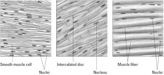
Today we talk about tissues in the human body. Muscle tissue is classified in three ways based on the tissue’s function, shape, and structure:

Smooth muscle tissue: So-called because it doesn’t have the cross-striations typical
of other kinds of muscle, the spindle-shaped fibers of smooth muscle tissue
do have faint longitudinal striping. This muscle tissue forms into sheets and
makes up the walls of hollow organs such as the stomach, intestines, and bladder.
The tissue’s involuntary movements are relatively slow, so contractions last
longer than those of other muscle tissue, and fatigue is rare. Each fiber is about
6 microns in diameter and can vary from 15 microns to 500 microns long. If
arranged in a circle inside an organ, contraction constricts the cavity inside the
organ. If arranged lengthwise, contraction of smooth muscle tissue shortens
the organ.

Cardiac muscle tissue: Found only in the heart, cardiac muscle fibers are
branched, cross-striated, feature one central nucleus, and move through involuntary
control. An electron microscope view of the tissue shows separate fibers
tightly pressed against each other, forming cellular junctions called intercalated
discs that look like tiny, dark-colored plates. Some experts believe intercalated
discs are not cellular junctions but rather special structures that help move an
electrical impulse throughout the heart.

Skeletal muscle tissue:
This is the tissue that most people think of as muscle.
It’s the only muscle subject to voluntary control through the central nervous
system. Its long, striated cylindrical fibers contract quickly but tire just as fast.
Skeletal muscle, which is also what’s considered meat in animals, is 20 percent
protein, 75 percent water, and 5 percent organic and inorganic materials. Each
multinucleated fiber is encased in a thin, transparent membrane called a sarcolemma
that receives and conducts stimuli. The fibers, which vary from 10
microns to 100 microns in diameter and up to 4 centimeters in length, are subdivided
lengthwise into tiny myofibrils roughly 1 micron in diameter that are suspended
in the cell’s sarcoplasm.

Now you know about tissues in the human body


tissues of the human body
types of tissues in human body
tissue in human body
how many tissues are in the human body

Monday, 9 July 2012

Human being digestive system


Before jumping into a discussion on the alimentary tract, we need to review some basic
terms.


Ingestion: Taking in food

Digestion: Changing the composition of food — splitting large molecules into smaller
ones — to make it usable by the cells

Deglutition: Swallowing, or moving food from the mouth to the stomach

Absorption: Occurs when digested food moves through the intestinal wall and into
the blood

Egestion: Eliminating waste materials or undigested foods at the lower end of the
digestive tract; also known as defecation


The alimentary tract develops early on in a growing embryo. The primitive gut, or archenteron,
develops from the endoderm (inner germinal layer) during the third week after conception,
a stage during which the embryo is known as a gastrula. At the anterior end (head end),
the oral cavity, nasal passages, and salivary glands develop from a small depression called
a stomodaeum in the ectoderm (outer germinal layer). The anal and urogenital structures
develop at the opposite, or posterior, end from a depression in the ectoderm called the
proctodaeum. In other words, the digestive tract develops from an endodermal tube with
ectoderm at each end.

Whereas the respiratory tract is a two-way street — oxygen flows in and carbon dioxide flows
out — the digestive tract is designed to have a one-way flow (although when you’re sick or
your body detects something bad in the food you’ve eaten, what goes down sometimes comes
back up).


Mouth Pharynx Esophagus Stomach Small intestine Large intestine

When you swallow food, it’s mixed with digestive enzymes in both saliva and stomach
acids. Circular muscles on the inside of the tract and long muscles along the outside of
the tract keep the material moving right through defecation at the end of the line.


Search terms:

process digestion human beings
digestive system of human body
digestive system of man


Explain the process of breathing


1 Breathing: The technical term is pulmonary ventilation, or the movement of air into
and out of the lungs. (Breathing is also called inspiration and expiration.)

2 Exchanging gases: This takes place between the alveolar cells in the lungs, the blood,
and the body’s cells in two ways:

   • Pulmonary, or external, respiration: The exchange in the lungs when blood
gains oxygen and loses carbon dioxide, transforming it from venous blood into
arterial blood

   • Systemic, or internal, respiration: The exchange within systemic capillaries
when the blood releases some of its oxygen and collects carbon dioxide from the
tissues

3 Adult breathing rate: About 12 to 20 times per minute.

4 Anoxia: Oxygen deficiency in which the cells either don’t have or can’t utilize
sufficient oxygen to perform normal functions.

5 Asphyxia: Lack of oxygen with an increase in carbon dioxide in the blood and
tissues; accompanied by a feeling of suffocation leading to coma.

6 Expiration or exhalation: The diaphragm returns to its domed shape as the
muscle fibers relax, via elastic recoil of the lungs and tissues lining the thoracic
cavity, the external intercostal muscles relax, and the internal intercostal muscles
contract. This movement pulls the ribs back into place, decreasing the volume of
the thoracic cavity and increasing pressure, forcing air out of the lungs.

7 Hypoxia: Low oxygen content in the inspired air.

8 Inspiration or inhalation: When the muscles of the diaphragm contract, its
dome shape flattens; simultaneously, the contraction of the external intercostal
muscles pulls the ribs upward and increases the volume of the thoracic cavity,
decreasing the intra-alveolar pressure. The pressure difference between the
atmosphere and the lungs diffuses air into the respiratory tract.

9 Lung capacity: The vital capacity plus the residual air.

10 Mediastinum: The region between the lungs extending from the sternum ventrally
(at the front) to the thoracic vertebrae dorsally (at the back), and superiorly
(top) from the entrance of the thoracic cavity to the diaphragm inferiorly
(at the bottom).

 11 Minimal air: The volume of air in the lungs when they’re completely collapsed
(150 cubic centimeters in an adult).

12 Phrenic nerve: The nerve that innervates (stimulates) the diaphragm.

13 Residual air: The volume of air remaining in the lungs after the most forceful
expiration (1,200 cubic centimeters in an adult).

14 Respiratory centers: Nerve centers for regulating breathing located in the
medulla oblongata, or brain stem. The centers are influenced by the amount of
carbon dioxide in the blood.

15 Tidal air: The volume of air inspired and expired in the resting state (500 cubic
centimeters in an adult).

16 Vital capacity: The volume of air moved by the most forceful expiration after a
maximum inspiration. It represents the total moveable air in the lungs (4,600
cubic centimeters in an adult).

Here’s what happens as you breathe in and out . Red blood cells use a
pigment called hemoglobin to carry oxygen and carbon dioxide throughout the body
through the circulatory system  Hemoglobin bonds loosely with oxygen, or O2, to
carry it throughout the body; the bonded hemoglobin is called oxyhemoglobin.

After hemoglobin releases its oxygen molecules, it picks up carbon dioxide, or CO2,
to deliver to the lungs for exhalation. The freshly bonded hemoglobin becomes
carbohemoglobin


Monday, 2 July 2012

Blood vessels in human body

The blood vessels in human body,Blood vessels come in three varieties

1 Arteries carry blood away from the heart. The largest artery is the aorta. Small
ones are called arterioles, and microscopically small ones are called metarterioles.

2 Veins carry blood toward the heart; all veins except the pulmonary veins contain
deoxygenated blood. Small ones are called venules, and large venous spaces are
called sinuses.

3 Microscopically small capillaries carry blood from arterioles to venules, but
sometimes tiny spaces in the liver and elsewhere called sinusoids replace
capillaries.

The walls of arteries and veins have three layers: the outermost tunica externa (sometimes
called tunica adventitia) composed of white fibrous connective tissue, a central
“active” layer called the tunica media composed of smooth muscle fibers and yellow
elastic fibers, and an inner layer called the tunica intima made up of endothelium that
aids in preventing blood coagulation by reducing the resistance of blood flow. Arterial
walls are very strong, thick, and very elastic to withstand the great pressure to which
the arteries are subjected. Arteries have no valves.

There are two types of arteries: elastic and muscular. In elastic arteries, found primarily
near the heart, the tunica media is composed of yellow elastic fibers that stretch
with each systole and recoil during diastole; essentially they act as shock absorbers to
smooth out blood flow. In muscular arteries, the tunica media consists primarily of
smooth muscle fibers that are active in blood flow and distribution of blood. The
larger blood vessels have smaller blood vessels, the vasa vasorum, that carry nourishment
to the vessel wall.

While larger in diameter than arteries, veins have thinner walls and are less distensible
and elastic. Veins that carry blood against the force of gravity, such as those in the legs
and feet, contain valves to prevent backsliding into the capillaries. Normally the blood
that veins are returning to the heart is unoxygenated (contains carbon dioxide); the
one exception is the pulmonary vein, which returns oxygenated blood to the heart
from the lungs.

Capillaries are breathtakingly tiny and capable of forming vast networks, or capillary
beds. Their walls are a single layer of squamous endothelial cells. Precapillary
sphincters take the place of valves to regulate blood flow. All exchange occurs at
the capillaries.

Blood from the digestive tract takes a detour through the hepatic portal vein to
the liver before continuing on to the heart. Called the hepatic portal system, this
circuitous route helps regulate the amount of glucose circulating in the bloodstream


What Is Human Anatomy?

Anatomy means the study of structure and human anatomy means the study of structure of human beings. It is one of the three basic medical sciences, which are taught to medical students who are to follow a career related to hospitals.

Human anatomy is purely related to the study of structure. It is not concerned with the study of functions of various parts of human body. In fact, there is another basic medical science, known as Physiology, which is concerned with the study of the function of various parts of human body. Anatomy just describes the structural details.

Yes, it is a fact that structure and function are very much inter-related and one cannot be understood without the other but a distinction has to be made because of the level of details in both fields. The details of human structure are so vast that they cannot be studied along with the vast details of human functions. That is why the study of function and structure is differentiated into two different branches of medical science.

It can be divided into three major categories.

1) Gross anatomy (macroscopic anatomy)
2) Microscopic anatomy (Histology)
3) Basic anatomy

Gross Anatomy: It deals with the study of macroscopic details of human structure. It is not concerned with fine microscopic structural details of human body and is studied with naked eye. It has two approaches of study: Systemic approach and regional approach. In systemic approach, the human body is considered to be composed of different organs systems while in regional approach, human body is considered to be composed of different regions.

Microscopic anatomy: It deals with the study of microscopic details of various structures of human body. Microscopic anatomy depends on an important instrument known as the microscope.
Basic anatomy: It is sometimes not considered as a major subdivision of human anatomy, however, it is very important for medical students who are new to the concepts of anatomy. Basic anatomy explains all the basic concepts of human anatomy so that the different structural arrangements of these basic components can be understood properly.
For more information, please visit the following website where all subdivisions of human anatomy including the body systems are explained in a smart way that makes the learning process very easy.
Human Anatomy
Article Source: http://EzineArticles.com/?expert=Ahsan_Doct_Iqbal


Article Source: http://EzineArticles.com/5885510

Study Anatomy

Study Anatomy

There are many different reasons for wanting to study anatomy. Maybe you want to learn human anatomy as a part of an educational program. Or, maybe you need to learn how the human body functions in order to further your career. Amazingly, you might just want to learn about the human body for personal reasons. Whether your reason is academic, business or personal in nature, learning about the human body is very intense, but rewarding.

Definitions

There are many technical and difficult definitions of human anatomy, but basically it is the study of body parts. Many people also learn physiology when they learn anatomy. Physiology explains how the body parts that you learned about in anatomy actually function. In order to understand how physiology works you have to also understand anatomy. It is for this reason that anatomy and physiology are usually taught together at most medical programs.

Considerations

Learning about the human body takes time and should be treated as a serious task. It is the foundation for learning about different facets of the medical profession. Depending on where you intend to specialize, anatomy training involves much more than just reading textbooks.
More advanced training goes beyond textbooks and may involve the use of learning tools such as graphic medical diagrams, anatomy photos and even human cadavers. If you are not comfortable about or strong enough to learn about the human body and its tissues and fluids, then the medical profession may not be a good career choice for you.

Misconceptions

Many people have the misconception that it is too hard to study anatomy. It is a challenge, but it can be done with the right tools. It has often been said that the average anatomy and physiology course is used to get rid of inadequate students that don't possess either the capacity to learn or the stomach to learn about the human body. Although this logic may not apply to you, it is up to the individual as to how and if you are going to tackle and learn anatomy.
Just like with any other challenge, find a strategy for accomplishing the task and stick with it. Human anatomy study guides will help you accomplish your goals. Contrary to belief, you can't rely on riddles, nursery rhymes and other memorization games to get you through anatomy and physiology courses.

Tips

Learning anatomy involves covering a lot of material. In order to be successful, you must have advanced study skills. You will not be able to get by and pass anatomy courses without studying. If you are one of those people that can pass all of your classes without studying, unfortunately, you will not have the same luck with the average anatomy class. Now is the time to purchase one of the best human anatomy study guides that your budget will allow.

These are just a few things that will help you to tackle and study anatomy successfully. Understand that learning about the human body is very challenging. However, all medical professionals must be knowledgeable about the human body. The topic of human anatomy is very challenging, but it can be mastered with the right mindset and learning tools.
As a medical student one needs to Study Anatomy Guide for a good understanding of human skeleton visit my blog to learn more about it.
Article Source: http://EzineArticles.com/?expert=Kurtis_D_Britt


Conduction System Of Heart

"Conduction System Of Heart"
The mighty, nonstop heart keeps up its rhythm because of a carefully choreographed
dance of electrical impulses called the conduction system that has the power to produce
a spontaneous rhythm and conduct an electrical impulse. Four structures play
key roles in this dance — the sinoatrial node, atrioventricular node, atrioventricular
bundle, and Purkinje fibers.Each is formed of highly tuned modified cardiac muscle.
Rather than both contracting and conducting impulses as other cardiac muscle does,
these structures specialize in conduction alone, setting the pace for the rest of the heart.
Following is a bit more information about each one:

1 Sinoatrial node: This node really is the pacemaker of the heart. Located at the
junction of the superior vena cava and the right atrium, this small knot, or mass,
of specialized heart muscle initiates an electrical impulse that moves over the
musculature of both atria, causing atrial walls to contract simultaneously and
emptying blood into both ventricles. It’s also called the S-A node, sinoauricular
node, and sinus node.

2 Atrioventricular node: The impulse that starts in the S-A node moves to this mass
of modified cardiac tissue that’s located in the septal wall of the right atrium. Also
called the A-V node, it directs the impulse to the A-V bundles in the septum.

3 Atrioventricular bundle: From the A-V node, the impulse moves into the atrioventricular
bundle, also known as the A-V bundle or bundle of His (pronounced
“hiss”). The bundle breaks into two branches that extend down the sides of the
interventricular septum under the endocardium to the heart’s apex.

4 Purkinje fibers: At the apex, the bundles break up into terminal conducting fibers,
or Purkinje fibers, and merge with the muscular inner walls of the ventricles. The
pulse then stimulates ventricular contraction that begins at the apex and moves
toward the base of the heart, forcing blood toward the aorta and pulmonary artery.

One of the best ways to detect cardiac tissue under a microscope is to look for undulating
double membranes called intercalated discs separating adjacent cardiac muscle
fibers. Gap junctions in the discs permit ions to pass between the cells, spreading the
action potential of the electrical impulse and synchronizing cardiac muscle contractions.
Potential problems include fibrillation, a breakdown in rhythm or propagation of
the impulses that causes individual fibers to act independently, and heart block, an
interruption that causes the atria and ventricles to take on their own rates of contraction.
Usually the atria contract faster than the ventricles.

A healthy heart makes a “lub-dub” sound as it beats. The first sound (the “lub”) is
heard most clearly near the apex of the heart and comes at the beginning of ventricular
systole (the closing of the atrioventricular valves and opening of the semilunar
valves). It’s lower in pitch and longer in duration than the second sound (the “dub”),
heard most clearly over the second rib, which results from the semilunar valves closing
during ventricular diastole. Defects in the valves can cause turbulence or regurgitation
of blood that can be heard through a stethoscope. Called murmurs, these sounds
indicate imperfect closure of one or more valves.


search terms:
conducting system of heart
conduction system disease
heart conducting system
conducting system of the heart
electrical conduction system of the heart

What is the function of liver

"What is the function of liver"
The largest gland in the body, the liver is divided into a large right lobe and a small left
lobe by the falciform ligament, another peritoneal fold. Two smaller lobes — the
quadrate and caudate lobes — are found on the lower (inferior) and back (posterior)
sides of the right lobe. The quadrate lobe surrounds and cushions the gallbladder, a
pear-shaped structure that stores and concentrates bile, which it empties periodically
through the cystic duct to the common bile duct and on into the duodenum during
digestion. Bile aids in the digestion and absorption of fats; it consists of bile pigments,
bile salts, and cholesterol.

The liver secretes diluted bile through the hepatic ducts into the cystic duct and on
into the gallbladder. Liver tissue is made up of rows of cuboidal cells separated by
microscopic blood spaces called sinusoids. Blood from the interlobular veins and arteries
circulates through the sinusoids with food and oxygen for the liver cells, picking up
materials along the way. The blood then enters the intralobular veins, which carry it to
the sublobular veins, which empty into the hepatic vein, which leads to the inferior
vena cava. Bile secreted from the liver cells is carried by biliary canaliculi (bile capillaries)
to the bile ducts and then to the hepatic ducts.

Considering the number of vital roles the liver plays, the complexity of that process
isn’t too surprising. Among the liver’s various functions are

1 Production of blood plasma proteins including albumin, antibodies to fend off
disease, a blood anticoagulant called heparin that prevents clotting, and bile pigments
from red blood cells, the yellow pigment bilirubin, and the green bile pigment
biliverdin

2 Storage of vitamins and minerals as well as glucose in the form of glycogen

3 Conversion and utilization through enzyme activity of fats, carbohydrates, and
proteins

4 Filtering and removal of nonfunctioning red blood cells, toxins (isolated by
Kupffer cells in the liver) and waste products from amino acid breakdown, such
as urea and ammonia

 Unfortunately, a number of serious diseases can damage the liver. The hepatitis virus
inflames the gland, and cirrhosis caused by repeated toxic injury (often through alcohol
or other substance abuse) destroys Kupffer cells and replaces them with scar
tissue. Also, painful gallstones can develop when cholesterol clumps together to form
a center around which the gallstone can form.


Terms
What is the function of liver

Wednesday, 20 June 2012

Human anatomy of head and neck

The skull is made up of 19 bones, 12 of which are pairs. You
can see most of these bones in the illustration. Where the
bones join is called a suture. Little fingers of bone interdigitate
with adjoining little fingers to make the joining solid.
The bones shown are: the frontal bone, the nasal bones, the
lacrimal bones, the ethmoid bones, the sphenoid bones, the
zygomatic bones, the maxilla, the mandible, the parietal
bones, the temporal bones, the occipital bone, the palatine
bones (not seen, since they are inside the orbit of the eye),
and the vomer (not seen, since it is inside the nasal cavity).

There are two bones that you cannot see in the illustration,
the palatine and the vomer. The palatine bones are paired
and are buried deep in the skull behind the nose. They make
up the rear part of the palate, part of the base of the nasal
cavity, and a small part of the floor or the orbit. The vomer is
a thin bone which forms part of the nasal septum separating
the two sides of the nasal cavity.

Lacrimal, meaning tear producing, is from the Latin lachrymal,
meaning a small vase, of the kind found in ancient
Roman sepulchers that was used for collecting tears shed in

mourning.(The lacrimal bone forms half of the receptacle,
which holds the lacrimal sac, a structure that receives the
tears and directs them into the nasal cavity. That explains
why we blow our noses in cold weather, or when we cry, we
are blowing out the tears that have drained into the cavity.
The other half of the receptacle for the lacrimal sac is made
from the frontal process of the maxilla.) Ethmoid, so-named
because it is full of holes, is from the Greek ethmo and oiedes,
meaning “formed like a strainer.” Sphenoid is from the Greek
spheno and eidos together meaning wedge-shaped. Zygomatic
or zygoma comes from the Greek zygon, which means yoke,
the kind used to harness oxen. Maxilla is from the Latin mala
meaning jaw, particularly the upper jaw. Mandible derives
from the Latin mandibula, which stems from mandare meaning
to chew and pertains particularly to the lower jaw, which
has most of the chewing motion. Parietal is from the Latin
paries, parietes, meaning “walls of a hollow cavity.” Temporal
indicates the temple, from the Latin tempora, meaning “temple,
the right place, the fatal spot..”(As well as indicating a
place on the skull where death can easily be afflicted, this
word coveys a sense of reverence for life.) Occipital is from
the Latin occiput, meaning “the back of the head.” Palatine is
from the Latin palatum, meaning the hard palate and is the
base for the words palatable and palliative. Vomer is the Latin
word for plowshare.

Search term
Human anatomy of head and neck


Tuesday, 19 June 2012

what is myology? The defination of myology

The study of muscles is called myology after the Greek word mys, which means “mouse.”
Muscles perform a number of functions vital to maintaining life, including

Movement: Skeletal muscles (those attached to bones) convert chemical energy into
mechanical work, producing movement ranging from finger tapping to a swift kick of a
ball by contracting, or shortening. Reflex muscle reactions protect your fingers when
you put them too close to a fire and startle you into watchfulness when an unexpected
noise sounds. Many purposeful movements require several sets, or groups, of muscles
to work in unison.

Vital functions: Without muscle activity, you die. Muscles are doing their job when
your heart beats, when your blood vessels constrict, and when your intestines squeeze
food along your digestive tract in peristalsis.

Antigravity: Perhaps that’s overstating it, but muscles do make it possible for you to
stand and move about in spite of gravity’s ceaseless pull. Did your mother tell you to
improve your posture? Just think how bad it would be without any muscles!

Heat generation: You shiver when you’re cold and stamp your feet and jog in place
when you need to warm up. That’s because chemical reactions in muscles result in
heat, helping to maintain the body’s temperature.

Keep the body together: Muscles are the warp and woof of your body’s structure,
binding one part to another.

As you may remember from studying tissues, muscle cells — called fibers — are some
of the longest in the body. Fibers are held together by connective tissue and enclosed
in a fibrous sheath called fascia. Some muscle fibers contract rapidly, whereas others
move at a leisurely pace. Generally speaking, however, the smaller the structure to be
moved, the faster the muscle action. Exercise can increase the thickness of muscle
fibers, but it doesn’t make new fibers. Skeletal muscles have a rich vascular supply
that dilates during exercise to give the working muscle the extra oxygen it needs to
keep going.

Two processes are central to muscle development in the developing embryo: myogenesis,
during which muscle tissue is formed; and morphogenesis, when the muscles form
into internal organs. By the eighth week of gestation, a fetus is capable of coordinated
movement.

Search terms;
what is myology
what the myology
mylogy
defination of myology

Arthrology, The Defination of Arthrology

Arthrology, which stems from the ancient Greek word arthros (meaning “jointed”), is
the study of those structures that hold bones together, allowing them to move to varying
degrees — or fixing them in place — depending on the design and function of the
joint. The term articulation, or joint, applies to any union of bones, whether it moves
freely or not at all.

Inside some joints, such as knees and elbows, are fluid-filled sacs called bursae that
help reduce friction between tendons and bones; inflammation in these sacs is called
bursitis. Some joints are stabilized by connective tissue called ligaments that range
from bundles of collagenous fibers that restrict movement and hold a joint in place to
elastic fibers that can repeatedly stretch and return to their original shapes.

The three types of joints are as follows:


Fibrous: Fibrous tissue rigidly joins the bones in a form of articulation called
synarthrosis, which is characterized by no movement at all. The sutures of the
skull are fibrous joints.

Cartilaginous: This type of joint is found in two forms:

• Synchondrosis articulation involves rigid cartilage that allows no movement,
such as the joint between the ribs, costal cartilage, and sternum.
• Symphysis joints occur where cartilage fuses bones in such a way that
pressure can cause slight movement, called amphiarthrosis. Examples
include the intervertebral discs and the symphysis pubis.

Synovial: Also known as diarthrosis, or freely moving, joints, this type of articulation
involves a synovial cavity, which contains articular fluid secreted from the
synovial membrane to lubricate the opposing surfaces of bone. The synovial
membrane is covered by a fibrous joint capsule layer that’s continuous with the
periosteum of the bone. Ligaments surrounding the joint strengthen the capsule
and hold the bones in place, preventing dislocation. In some synovial joints,
such as the knee, fibrous connective tissue called meniscus develops in the
cavity, dividing it into two parts. In the knee, this meniscus stabilizes the joint
and acts as a shock absorber.

What is muscle tissue made up of

The question in your minds about what is muscle tissue made up of.in histology you should know that muscle tissue is made up of fibers known as myocytes. The cytoplasm within the fibers
is called sarcoplasm, and within that sarcoplasm are minute myofibrils that contain the
protein filaments actin and myosin. These filaments slide past each other during a
muscle contraction, shortening the fiber.

Following are the three types of muscle tissue

Smooth muscle tissue: This type of tissue contracts without conscious control.
Made up of spindle-shaped fibers with large, centrally located nuclei, it’s found in
the walls of internal organs, or viscera. Smooth muscle gets its name from the
fact that, unlike other muscle tissue types, it is not striated.

Cardiac muscle tissue: Also known as myocardium, cardiac muscle tissue is
made of branching fibers, each with a central nucleus and alternating light and
dark striations. Between the fibers are dark structures called intercalated discs.
As with smooth muscle, cardiac muscle tissue contractions occur through the
autonomic nervous system.

Skeletal, or striated, muscle tissue: Biceps, triceps, pecs — these are the muscles
that bodybuilders focus on. As the name implies, skeletal muscles attach to
the skeleton and are used throughout the central nervous system for movement.
Muscle fibers are cylindrical with several nuclei in each cell (which makes them
multinucleated) and cross-striations throughout.


What is muscle tissue made up of


“ Take A Journey Inside The Mysterious
      World Of The Human Body ... ”

This heavily illustrated self-teaching course
gives you everything you need to -
CLICK HERE


Monday, 18 June 2012

Get under your skin definition

Perhaps because of its unique job of both protecting the outer body and lining internal
organs, epithelial tissue comes in more varieties than any other tissue.

Epithelial tissues, which generally are arranged in sheets or tubes of tightly-packed cells,
always have a free, or apical, surface that can be exposed to the air or to fluid. That free surface
also can be covered by additional layers of epithelial tissue. But whether it’s layered or
not, each epithelial cell has polarity, and all but one side of the cell is
tucked snugly against neighboring cells. The apical side sometimes has cytoplasmic projections
such as cilia, hair-like growths that can move material over the cell’s surface, or
microvilli, finger-like projections that increase the cell’s surface area for absorption. Opposite
the apical side is the basal side (think basement), which typically attaches to some kind of
connective tissue.

Epithelial tissue serves several key functions, including the following:

Protection: Skin protects vulnerable structures or tissues deeper in the body.

Barrier: Epithelial tissues prevent foreign materials from getting inside the body.

Sensation: Sensory nerve endings embedded in epithelial tissue connect the
body with outside stimuli.

Secretion: Epithelial tissue in glands can be specialized to secrete enzymes, hormones,
and fluids.

Single-layer epithelial tissue is classified as simple. Tissue with more than one layer is
called stratified. Epithelial tissues also can be classified according to shape: Squamous
is a thin, flat cell; cuboidal is, as the name implies, equal in height and width and
shaped like a cube; and columnar cells are taller than they are wide.